Materials
- Clear plastic bottle, such as a 2 liter bottle
- Cork stopper that will fit the mouth of the bottle. This cork should have a hole through it.
- Small tube that will fit the hole in the cork
- Large balloon
- A large piece of broken balloon. A used party balloon works well for this.
- Tape -Wide clear packing tape works the best.
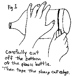 (Fig.1)
Carefully cut the bottom off of the plastic bottle. Put tape all around
the bottom, covering the sharp edge. This is to protect the balloon that
you will attach later. Insert the tube into the cork so that the tube
sticks out on both ends.
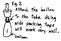 (Fig
2) Attach the balloon to the tube using tape. Make sure it is completely
sealed. To test it, try blowing up the balloon.
The balloon represents a lung. There are no muscles in the lungs.
*If you can get the mouth of the balloon over the mouth of the bottle, then you will not need the cork.
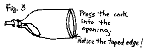 (Fig.
3) Put the cork into the mouth of the plastic bottle. Make sure it is
tight.
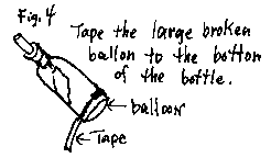
(Fig. 4) Tape the large broken balloon to the bottom of the plastic bottle. Make sure it is air tight!
This balloon represents the large muscle that is beneath the lungs (the diaphragm).
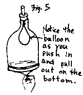
(Fig. 5) Push and pull on the balloon (slowly) to see how it works. If you have trouble catching hold of the balloon, put a small piece of tape on the center of the balloon to make a handle.
What happens to the balloon (lung)?
Text Source (modified): 39 Easy Animal Biology Experiments by Robert Wood
Gas exchange in the lungs. To supply oxygen to the blood and remove carbon dioxide from it, the lungs need to draw in fresh gas and expel stale gas. Fresh gas is drawn in when the diaphragm and other muscles in the chest wall contract. This action--called inspiration or inhalation--makes the chest volume larger and causes the lungs to expand. The expansion lowers the pressure in the lungs, and air from the atmosphere flows in. When the muscles relax, the lungs return to a smaller volume, and gas flows out into the atmosphere. This action is called expiration or exhalation.
Diaphragm, pronounced DY uh fram, the large muscle attached to the lower ribs, separates the chest from the abdomen. Only human beings and other mammals have complete diaphragms. The diaphragm is the chief muscle used in breathing. It is shaped like a dome.
When a person takes a breath, the diaphragm contracts and moves downward. This increases the space in the chest. At the same time, muscles attached to the ribs cause the ribs to move outward. This expands the chest, and together with the downward motion of the diaphragm, creates a slight vacuum in the chest. The vacuum causes air to enter the lungs through the windpipe. This action is called inspiration or inhalation.
During expiration, also called exhalation, air moves out of the lungs as the diaphragm and rib muscles relax. When a person breathes normally, expiration is passive and muscles do no work. The expanded lung contains elastic fibers that were stretched during inspiration. This elastic tissue behaves like stretched rubber bands, causing the lung to contract like a collapsing balloon. This forces air out of the chest. The lung gets smaller until it reaches the size at which the breath started. The lungs do not empty completely during expiration because the chest wall holds them in a partially expanded state. In hard breathing, as occurs during exercise, expiration is active. Another set of rib muscles helps to make the chest smaller. Muscles in the abdominal wall also contract to push the abdominal organs upward against the diaphragm, helping force air out of the lungs.
The phrenic nerve carries the electrical signals to the diaphragm that
stimulate it to contract. This nerve arises from the spinal cord high in
the neck and extends into the chest down to the diaphragm.
Back to Fig. 4
from World Book
January 2, 2000


 DonnaYoung.org has 62 more manuscript handwriting animations!
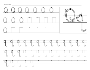 Handwriting practice worksheets in a font style that is similar to handwriting w/o tears.


